The Shanghai Soft X-ray Free-Electron Laser facility (SXFEL)
The Shanghai Soft X-ray Free-Electron Laser facility (SXFEL), located at the SSRF campus, is developed in two phases: the SXFEL test facility (SXFEL-TF) which started in 2014 and the SXFEL user facility (SXFEL-UF) which started in 2016. The test facility achieved its design goal and passed the national acceptance in Nov. 2020. The construction and commissioning of the user facility was finished in 2022. The user operation was started in Jan. 2023.
SXFEL is the first X-ray free-electron laser facility in China and one of the core platforms of the Zhangjiang photon science facility cluster, which firmly supports the construction of Zhangjiang Comprehensive National Science Center. The facility is 532 meters long in total, consisting of 1 injector, 1 main linac, 1 switchyard, 2 undulator lines, 2 beamlines and 5 experimental stations. SXFEL will provide the scientific users with cutting-edge research means such as ultrafast process observation, advanced structure analysis, high-resolution imaging, etc., and become the original innovation source and talent cradle of new discoveries in scientific research, new principles, new technologies, and new methods in the field of free-electron laser.

Milestones
 |  |  |  |  |
| 2014.12 SXFEL-TF started | 2016.5 SXFEL-TF civil construction finished | 2016.12 SXFEL-UF and SBP started | 2020.8 SXFEL-TF finished | 2023.1 SXFEL user operation started |
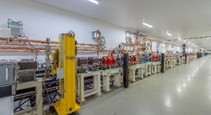 | Injector |
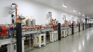 | Main Linac The main linac of SXFEL starts with a S-band accelerating section (L1) consisting of two 3m-long accelerating structures, and this is followed by an X-band accelerating structure called linearizer used for compensating the high order correlated energy spread before the first bunch compressor (BC1). After the bunch compressor, two high gradient C-band accelerating sections (L2 and L3) are employed to accelerate the electron to the required energy. The accelerating gradients for these S-band and C-band structures are set at 27 MV/m and 38 MV/m respectively. The linac can provide a high-quality electron beam with energy of 1.5 GeV, charge of 0.5 nC, peak current of about 700 A and normalized project emittance of about 1.5 mm·mrad. |
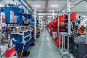 | Undulator A beam distribution switchyard lies downstream of the transport line after the linac, distributing the beam to two downstream undulator lines while maintaining the quality of the electron beam. Two undulator lines, a SASE line and a Seeding line, will produce FEL radiation with the shortest wavelength of about 2 nm and 3 nm, respectively. Two helical undulators located at the end of the seeding FEL line aiming to provide polarization-controllable, fully coherent radiation pulses. |
 | Beamline SXFEL facility includes two beamlines, delivering SASE and Seeding pulses respectively. The SASE beamline has two branches which can provide the focused pink and monochromatic beam for the endstations, e.g., CSI, TXS and UXS; It covers the photon energy 100~620eV, with the repetition rate 1~50Hz, single pulse intensity ~100μJ, bandwidth ~600meV, pulse length 50~500fs and focusing spot size at sample position ~3μm. The seeding beamline has two branches as well, which can provide the focused beam for the endstations AMO and VMI etc.; It covers the photon energy 50~410eV with the repetition rate 1~50Hz, single pulse energy ~50μJ, bandwidth 50~500meV, pulse length 50~500fs and focusing spot size at sample position ~10μm. Diagnostic tools, e.g. spectrometer, gas monitor detector, timing tool, coherence measurement device etc., have been developed to measure the spectrum, intensity, temporal profile etc. for SXFEL facility. |
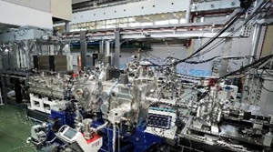 | Coherent Scattering and Imaging (CSI) Coherent Scattering and Imaging endstation is combined with advanced fluorescent super-resolution microscopic imaging technology, integrating the first X-ray free-electron laser and fluorescent super-resolution nano imaging system in the world. It can realize dynamic imaging of cell structure and function. It is characterized by using atomic XFEL coherent high brightness femtosecond pulses, combining coherent diffraction imaging to realize the pre damage imaging of the sample, and obtain the real structure information of the sample. It also covers cutting-edge research areas such as fine structure analysis of novel materials, multi-physical field in-situ imaging and X-ray - matter interaction. |
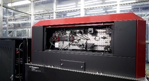 | Live-cell fluorescent Super-Resolution Microscope (SRM) The live-cell fluorescent super-resolution microscope endstation is to label the target structure with fluorescence probe, to illuminate the fluorescent sample with laser of suitable wavelength, and combine with four PI imaging technology, to realize single cell three-dimensional super-resolution imaging. It can be used to observe cell and tissue morphology, subcellular structure, etc. Further combined with X-ray imaging technology, it can realize the function of ultrastructure and fluorescence, in-situ composite imaging. |
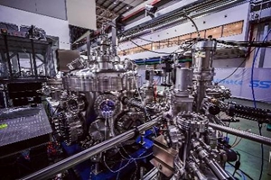 | Time-resolved X-ray Scattering (TXS) Using it, we can study the electronic states and lattice structures of various elements in materials, also the ultrafast dynamic changes within one billionth of a second. Utilizing this experimental station, we can study high-temperature superconductors, and study novel materials with strong correlation between electrons and electrons, between electrons and lattices and interaction. We can also study the ultrafast behavior of electrons in nonequilibrium state in quantum materials provides the most advanced material platform for the new type of quantum ultrafast devices. |
 | Ultrafast X-ray Spectroscopy for Chemistry (UXS) The ultrafast X-ray spectroscopy endstation is equipped with a grating-based X-ray spectrometer with a cross-bar optical design to resolve the x-ray energies and the time-delays at the same time. The optical pumping laser is irradiating normally on to the sample surface while the probing XFEL pulses incidental to the sample surface with a grazing angle of 2 degree. This cross-bar optical design encodes the time-delay between the two pulses onto the sample surface, which is imaged on to the detector. The Ultrafast X-ray Spectroscopy for Chemistry system can be used to study the ultrafast processes including chemical reaction kinetics, energy/charge-transfer process. |
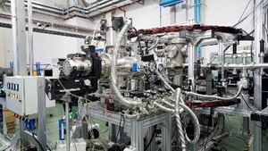 | Atomic, Molecular and Optical (AMO) The reaction microscope (REMI) at the Shanghai soft x-ray free electron laser for AMO science consists of a supersonic gas injection system, a spectrometer, and detectors with a data acquisition system. The pump-probe scheme will be provided with the synchronization of the external optical laser to the XFEL pulse. An Even-Lavie type high temperature pulsed valve will be implemented to further extend from gas sample to liquid or even volatile biomolecules in solid states. By measuring the time of flight and impact positions of ions and electrons on their corresponding detector, three-dimensional momentum vectors can be reconstructed in a coincidence manner. Momentum resolutions of ions and electrons with 0.11 a.u. can be achieved. |


 Copyright©2006.12 Shanghai Advanced Research Institute.