About
News and Events
Beamlines
- User Facilities
- Beamlines Directory
- BL08U1-A
- BL08U1-B
- BL09U
- BL13W1
- BL14W1
- BL14B1
- BL15U1
- BL16B1
- BL17U1
- BL01B1
- BL17B1
- BL18U1
- BL19U1
- BL19U2
Technology
- Accelerator Physics
- Accelerator Operations
- Radio Frequency
- Beam Instrumentation
- Control Systems
- Electronics & Detector
- Mechanical Engineering
- Vacuum
- Magnets
- Magnet Power Supplies
- Pulse Technique
- Cryogenics
- Front Ends
- Optics
User Information
Science and Publications

BL08U1-A Soft X-ray Spectromicroscopy Beamline
1. Introduction
X-ray spectromicroscopy is a technique for high brilliant tunable synchrotron radiation, which could combine the two important functions such as the sub-50 nm resolving ability in space determined by the zone plate, and the chemical distinguishing ability determined by the beamline monochromator via NEXAFS spectroscopy. The photon energy of BL08U1-A was range from 250 to 2000 eV, covering the K-edge of C, N, O, F, Na, Mg, Al, Si, and L edges of Cl, K, Ca, Fe, Cu, Zn. The study of these elements is very important in biological science, environmental science, polymer, and material science. In addition, the light source was finally decided by an elliptical polarized undulator, which would add more opportunities to study x-ray-polarization-dependent materials.
2. Beamline Layout
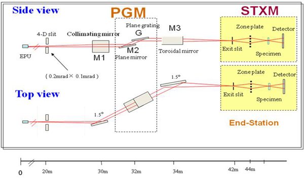
PGM- Plane Grating Monochromator STXM- Scanning Transmission X-ray Microscopy
3. Techniques
Ø Scanning transmission X-ray Microscopy and Spectroscopy (STXM)
Ø Transmitted Near Edge X-ray Absorption Spectroscopy (NEXAFS)
Ø Point spectrum scanning: to measure X-ray absorption near-edge spectra (XANES) at a point position of sample.
Ø Dual-energy contrast imaging:to quantitatively measure 2D spatial distribution of elements.
Ø Energy stacking method:to measure chemical composition and its spatial distribution, corresponding XANES of element.
Ø Total electron yield method (TEY):to measure XANES in a large sample area.
Ø Nano-CT
Ø CDI
4. Endstations
Scanning transmission X-ray Microscope (STXM)
A Fresnel zone plate is used to focus monochromatic X-rays to a small spot size (~50 nm). The sample is scanned through the focal point while detecting transmitted photons. While the technique is mainly bulk-sensitive, since samples tend to be very thin to allow adequate transmission in the soft X-ray region (smaller than 2micrometers), surface adsorbed species can often be detected by TEY.
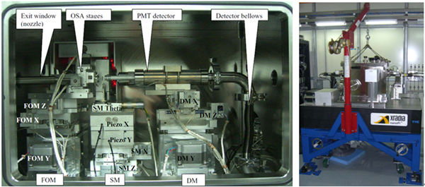
Detectors include the following parts:
Ø Photomultiplier tube (PMT)
Ø Photodiode (PD)
Ø Visible light microscope (VLM)
Ø CCD
5. Support LAB
Users can prepare the samples in the support lab. The followed instruments can be available:
Ø Visible light microscope: to observe the sample prepared.
Ø Slicing machine: to cut the sample into slices with thickness from 0.1mm to 10mm
Ø Ultrasonic cleaning machine
6. Beamline Specifications
|
Source |
Elliptically Polarized Undulator (EPU) |
|
Energy Range |
250-2000 eV |
|
EnergyResolution(ΔE/E) |
11000@244eV 2500@1560eV |
|
Photon Flux at sample |
8´107phs/s@244eV@200 mA 7´107phs/s@1840eV@200 mA |
|
Spatial resolution |
Better than 30 nm |
7. Applications
l Graphene-templated formation of ultrathin two-dimensional single crystal lepidocrocite nanosheets

Graphene-templated 2D single crystal lepidocrocite nanosheets (valance Fe3+, γ-FeOOH ) nanosheets were synthesized, it is an efficient catalyst for phenol treatment in wastewater. Most of the Fe element is transformed into lepidocrocite (γ-FeOOH, Fe (III) value) nanosheets. Energy & Environmental Science 2011, 4, 2035-2040.
l Metalloreductase Steap3 coordinates the regulation of iron homeostasis and inflammatory responses
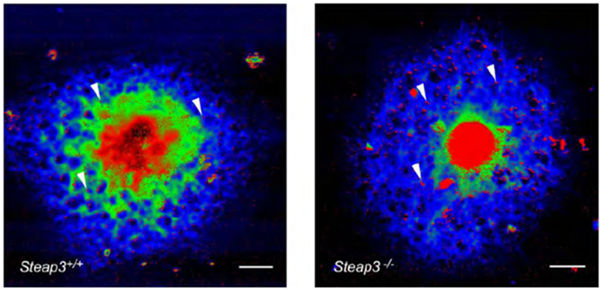
Steap3 is important in regulating both iron homeostasis and TLR4-mediated inflammatory responses in macrophages. STXM results proved that Steap 3 deficiency causes abnormal iron status and homeostasis, which leads to impaired TLR4-mediated inflammatory responses in macrophages. Following inflammatory stimuli, Steap3 depletion causes disregulated iron sequestration and distribution. Haematologica 2012, 97 (12): 1826-1835.
l Biotransformation of Ceria Nanoparticles in Cucumber Plants
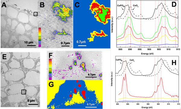
STXM imaging and Near edge X-ray absorption fine structure (XANES) spectra showed that Ce presented in the roots as CeO2 and CePO4 while in the shoots as CeO2 and cerium carboxylates. This is the first time to prove the transformation of CeO2 nanoparticles which can be one of important information on the biotoxicity of CeO2. ACS Nano 2012,6 (11): 9943-9950.
Director: Renzhong Tai, tairenzhong@sinap.ac.cn
Beamline director: Yong Wang, wangyong@sinap.ac.cn, +86-21-33933208
User contact: Lijuan Zhang, zhanglijuan@sinap.ac.cn, +86-21-33932087
Beamline: +86-21-33932080
 Copyright©2006.12 Shanghai Advanced Research Institute.