1. Introduction
The X-ray Photoemission Spectroscopy and Microscopy Beamline (Dreamline) provides a state-of-the-art experimental set-up to study the electronic band structure of novel complex materials and the surfaces and interfaces of solid-state materials by using Angle-Resolved Photoelectron Spectroscopies (ARPES) and Photoemission Electron Microscopies (PEEM). The beamline was built with two APPLE-II type undulators (LEID and HEID) and produces a broad energy range from 20 to 2000 eV with high resolution, variable polarization, and high flux, as well as the minimized higher orders harmonics for the low energy photons. Due to this unique Duo-undulator technique, Dreamline provides an ideal platform for investigating the materials that has distinct properties from the bulk to the surface.
2. Beamline Layout
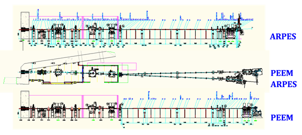
Beamline layout
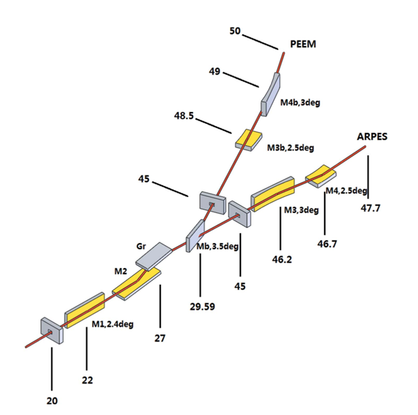
Optics
3.Techniques
ARPES End-station
· UVU ARPES
· Soft X-ray ARPES
· Resonant X-ray ARPES
· MBE
XPEEM End-station
· Bright & dark field LEEM
· MEM
· UV-PEEM
· X-ray PEEM
· LEED
· Spectroscopy
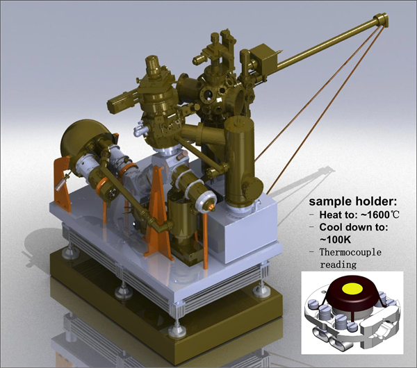
4. Endstations
ARPES End-station

The end-station is equipped with a DA30L analyzer with the updated deflection mode capable for taking Fermi surface with high efficiency, and a 6-axis cryogenic manipulator Carving? providing 3 translation and 3 rotation degree of freedom. This very solid and stable manipulator enables precise sample manipulating controlled by a sophisticate Labview-based software. The endstation also contains a load-lock with 6 parking position available, a transfer chamber providing Ar-sputtering, a preparation chamber equipped with a LEED and a K-cell, and a magnetic shielded main acquisition chamber. Besides, the endstation has a MBE, which can be used for in-situ growing and photoemission studies for Fe(TeSe) thin films.
The specification of the station is listed as below:
2 ?Eoptimal = 5 meV @ 20 eV , 21 meV @ 400 eV
2 Base temperature Tbase= 12 K (Carving) / 7 K (4-axis Manipulator)
2 Samples preparation: Ar-sputtering, annealing up to 1000℃,K-cell evaporators
2 In-situ Fe(TeSe) growth and characterization (MBE )
XPEEM End-station
The instrument allows to image samples using the photoelectric effect with very high spatial resolution, chemical and magnetic sensitivity. With an energy analyzer the excited photoelectrons can be energy-selected. In addition to illumination by X-rays, illumination by low energy electrons is possible. In this low energy electron microscopy (LEEM) mode additional contrast mechanisms are available.
The special/energy resolution data:
LEEM/PEEM: Field of view: 0.65-100um
2 Typical lateral resolution:
2 LEEM imaging: ~ 3nm
2 PEEM imaging: UV lamp: ~6nm
X ray: ~ 20nm
2 Energy resolution: ~0.15eV
2 Ion gun: sputter and clean sample surface
2 Femtosecond laser: 800nm and 400nm wavelength
2 Mercury lamp: photon 4. 9eV
2 Gas: O2, Ar, etc.
2 K-cell and E-beam evaporators: in situ growth and observation
5. Support Facilities
Glove Box: A Unilab Pro SP(1250/1000) glove box is available for preparing and storing air-sensitive samples
6. Beamline Specifications
|
Energy range |
20-200 eV(LEID),200-2000 eV(HEID) |
|
Resolving power |
35000 @ 867 eV |
|
Polarization |
Linear Horizontal, Linear Vertical,Circular Left/Right |
|
Flux on sample |
3.5×10¹¹ phs/s/0.01% BW @ 800eV |
|
Beam spot |
20 ?m×30 ?m |
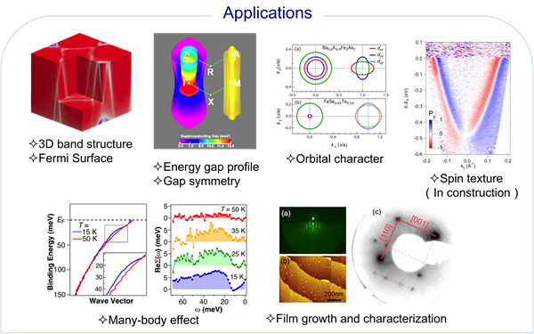
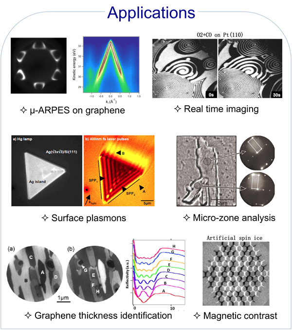
7. Applications
8. Contact
Yaobo Huang
huangyaobo@sinap.ac.cn
+86(0) 21 3393 3224

 Copyright©2006.12 Shanghai Advanced Research Institute.