Rotating the sample and imaging the sample by STXM at multiple angles, and then computerized tomographic reconstruction of the 2D images at each angle results in a 3D image of the sample.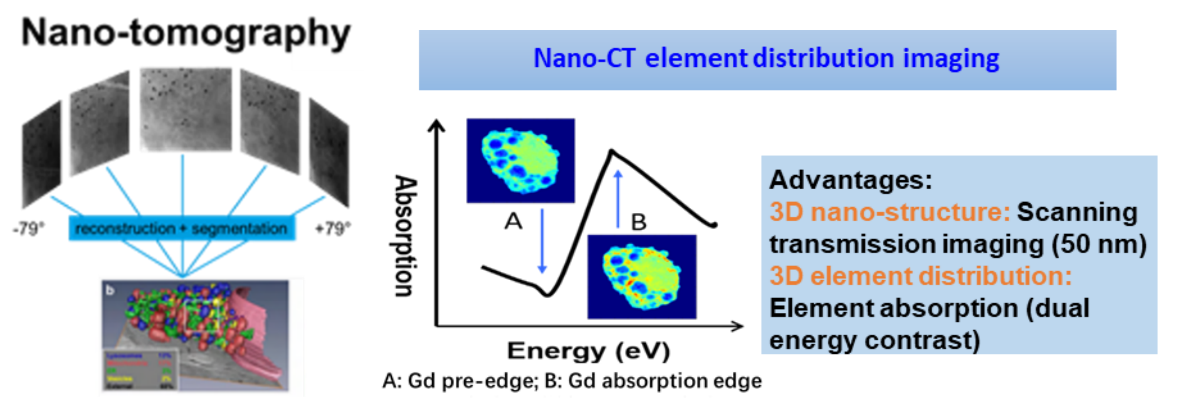
This beamline combines chemical element identification and 3D structural imaging of samples to realize characterization of sample 3D structures and element 3D distribution at nanoscale.
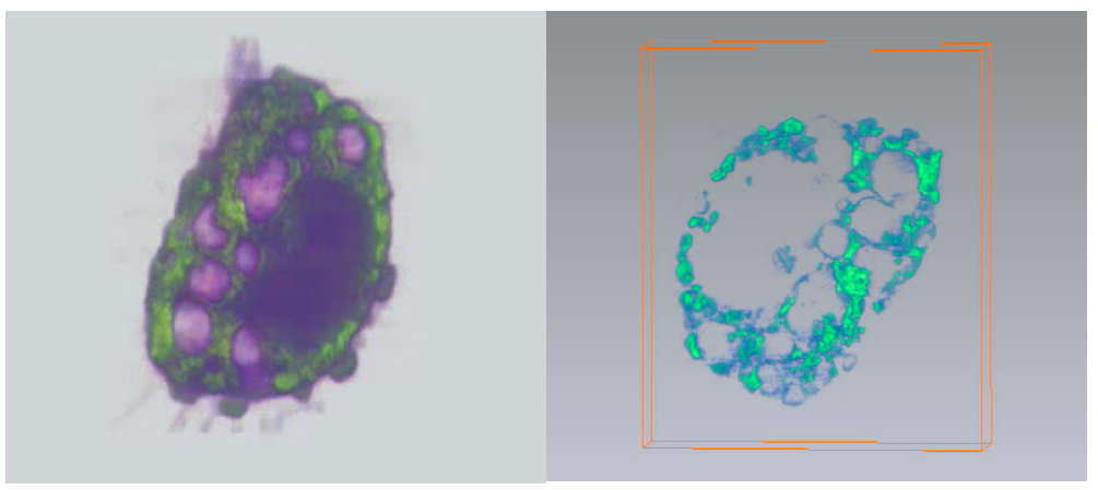
Recognition uptake and intracellular localization of metallofullerol Gd@C82(OH)22 by immune cells [IUCrJ 2018, 5, 141]
3D nano-imaging of an adherent immune cell (left) and 3D distribution of intracellular Gd elements (right).
附件下载: