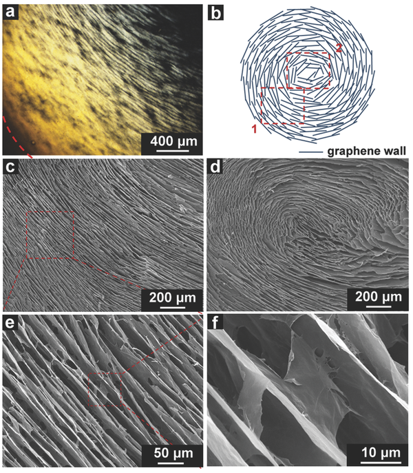
(a) POM images of a 3.5 mg mL−1 GO suspension containing 0.132 m KOH near the inner surface of glass tube (the dashed red arc indicates the isotopic glass wall). (b) Schematic illustration of the arrangement of graphene sheets in OGF (top view). (c–f) Scanning electron microscopy images of OGF with different magnifications. The images in panels (c) and (d) correspond to the area 1 and area 2 in scheme (b).
Links: DOI: 10.1002/adma.201504594