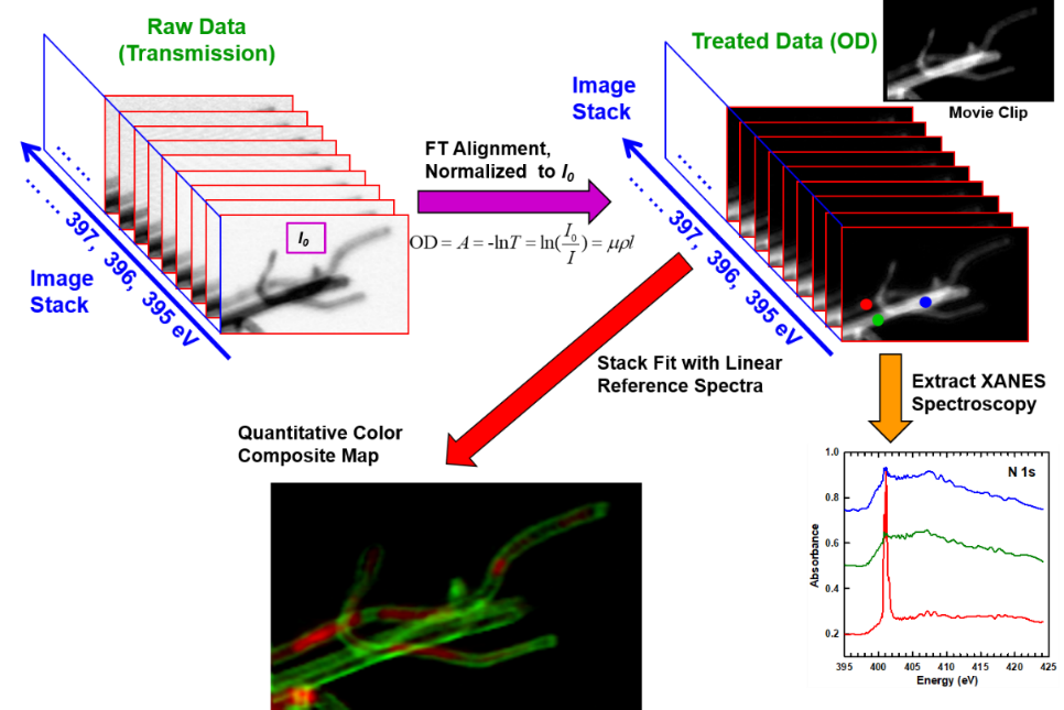
In this method, a series of 2D imaging of a sample at a series of energy points near the absorption edge of an element is performed. After alignment, normalization, PCA analysis, and reference spectrum fitting of these 2D images (image stack), the quantitative spatial distribution of the chemical states of the element and the corresponding absorption spectra can be obtained.
J. Phys. Chem. Lett. 2010, 1, 1709
Principle of energy stack imaging analysis method
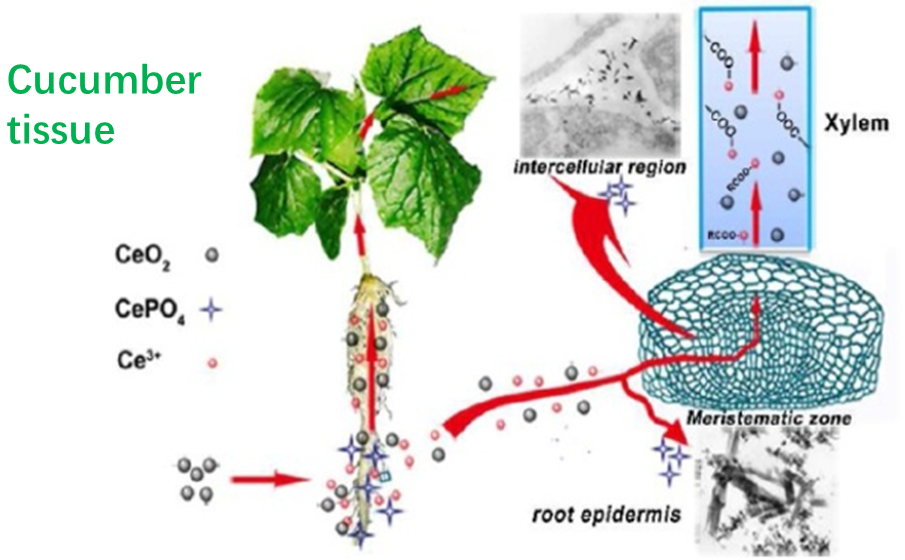
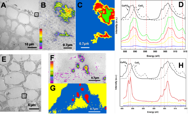
Biotransformation of cerium nanoparticles in cellular tissues. Stack imaging revealed that the chemical composition of the needle-like nanoclusters was cerium phosphate, confirming the transformation of cerium dioxide nanoparticles. This is the first time that cerium dioxide nanoclusters have been found and demonstrated to be able to be transformed in biological systems, providing important information for the evaluation of the ecotoxicology of cerium dioxide nanoclusters. [ACS Nano 2012, 6, 9943]
附件下载: